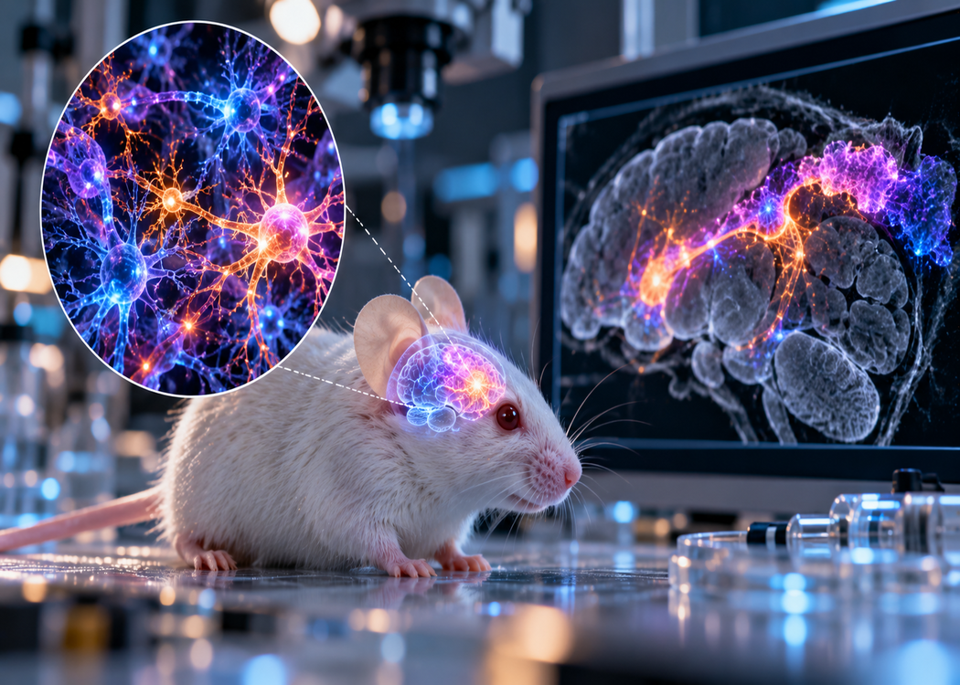
The experimental model could help scientists study developing human neurons within a living nervous system and investigate potential treatments for neurological disorders.
Scientists at Stanford Medicine have transplanted laboratory-grown human brain tissue into mice missing most of their cerebral cortex, allowing the tissue to grow and establish extensive connections with the animals’ nervous systems.
The study, published in Nature on September 16, introduces an experimental platform for investigating human brain development and disease. In its coverage, Nature described the results as the most extensive integration of human brain cells into an animal demonstrated so far.
The researchers used cortical organoids: three-dimensional clusters of tissue grown from human stem cells that reproduce some features of the developing cerebral cortex. These structures give scientists access to processes that are difficult to observe in living people, but laboratory cultures lack the full biological environment in which a brain normally develops.
Earlier transplantation experiments helped human neurons mature inside rodents. However, the animals’ existing brain tissue limited the space available for growth and competed with the grafts as connections formed.
To address that problem, the Stanford team genetically engineered mice with a major reduction in neurons in the neocortex and hippocampus. They then implanted human cortical organoids into the resulting cavity shortly after birth, creating what they call “xenocortical” mice.
Over the following months, the human tissue expanded and extended nerve fibres into the remaining mouse nervous system, including the spinal cord. Nature reported that the grafts filled more than 90% of the previously vacant space within two to three months. That figure refers to the space available for the graft, rather than the entire mouse brain.
The integration also involved functioning neural circuits. Electrical recordings and imaging revealed organised activity in the transplanted tissue resembling developing networks. Behavioural assessments identified differences in movement and coordination, but the findings do not demonstrate human-like intelligence or consciousness in the animals.
To explore the model’s research potential, the team examined how developing human cells responded to oxygen deprivation. The experiments produced cellular signs of injury and changes in gait, providing a possible route for studying injuries associated with oxygen shortages around birth. The researchers envision using the platform to investigate developmental disorders and evaluate candidate treatments; it is not itself a treatment for patients.
The work also makes ethical oversight central to the research. The study reports approval from Stanford’s stem-cell research oversight and animal-care committees, alongside monitoring of animal wellbeing and systematic testing for changes in cognitive and movement abilities.
The model’s promise lies in connecting observations of human cells with activity across a living nervous system. Establishing how faithfully those observations reflect human development will be essential to determining which scientific and therapeutic questions it can reliably answer.
Sources: