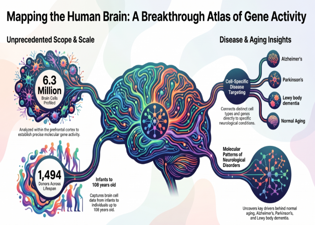
An atlas built from 1,494 donors reveals patterns linked to ageing and brain disorders, offering a detailed resource for investigating cellular vulnerability.
Scientists have created a vast atlas of gene activity in the human prefrontal cortex, examining more than 6.3 million cell nuclei from 1,494 deceased donors whose ages ranged from infancy to 108 years.
Developed through the PsychAD consortium, the resource allows researchers to investigate how different brain cell types vary across individuals, stages of life and neurological or psychiatric conditions. It focuses on the dorsolateral prefrontal cortex, a region involved in planning, decision-making and other complex cognitive functions.
A series of studies published in September in Nature and related journals uses the dataset to investigate healthy development, ageing and disease. The work brings multiple conditions into a shared analytical framework, helping researchers identify both overlapping molecular patterns and differences between disorders.
The researchers used single-nucleus RNA sequencing, which measures RNA within individual cell nuclei to assess gene activity. Examining these profiles separately allows scientists to distinguish changes in particular cell populations that could be obscured when tissue is analysed as a mixture. The resulting disease atlas distinguishes eight broad cell classes, 27 subclasses and 65 subtypes.
One major analysis compared eight categories of brain disorders, including Alzheimer’s disease, Parkinson’s disease, diffuse Lewy body disease, vascular dementia, frontotemporal dementia, tauopathy, schizophrenia and bipolar disorder, alongside neurotypical controls.
Researchers found shared gene-expression signatures involving basic cellular processes, including RNA processing and the placement of proteins within cells. After accounting for these widespread similarities, Alzheimer’s, Lewy body disease, vascular dementia and Parkinson’s showed particularly strong molecular overlap.
In Alzheimer’s disease, greater severity was associated with lower neuronal abundance and increased representation of immune and vascular cell populations. These findings draw attention to interactions among nerve cells, immune responses and blood vessels, rather than attributing the disease to a single cell type.
A separate lifespan study examined a subset of the resource: approximately 1.3 million nuclei from 284 neurotypical donors aged from birth to 97. This analysis provides a reference for understanding how gene activity changes during development and ageing, helping researchers distinguish age-associated patterns from those linked to disease.
The atlas has important limits. It examines one brain region using tissue collected after death, rather than following the same people throughout their lives. Associations between gene activity and disease therefore require further investigation to determine whether they reflect causes, consequences or responses to damage.
Its immediate value is as a research foundation. Scientists can use the resource to identify promising cell populations and molecular pathways for follow-up experiments. Over time, that work could help explain why certain cells are vulnerable—and guide efforts to develop more precisely targeted treatments.
Sources:
- Original disease study — Single-cell atlas of transcriptomic vulnerability across brain disorders
- Dataset paper — Population-scale cross-disorder atlas of the human prefrontal cortex at single-cell resolution
- PsychAD Consortium — Research findings and data resources
- Nature Portfolio — Detailed brain maps probe development, ageing, decline and disease